


Table of Contents
The oral cavity is always kept moist to prevent aberrations, ulcerations, and bacterial growth. This function is performed by the saliva secreted by the salivary glands. The largest salivary gland is the parotid gland which produces up to 50-60% of the total saliva produced by the glands of the oral cavity. These glands secrete their products when they get parasympathetic stimulation from the central nervous system. In the case of the parotid salivary gland, the parasympathetic supply is provided via the glossopharyngeal nerve through its parasympathetic fibers. In the following text, we shall discuss the origin, course, classification, and functional components of the glossopharyngeal nerve. We shall also discuss different nuclei related to it, its functions, its role in salivation and taste pathways, and different reflexes related to the glossopharyngeal nerve. Last but not least, we shall have a look at the diseases caused by damage to the glossopharyngeal nerve and the ways to check for its different lesions.
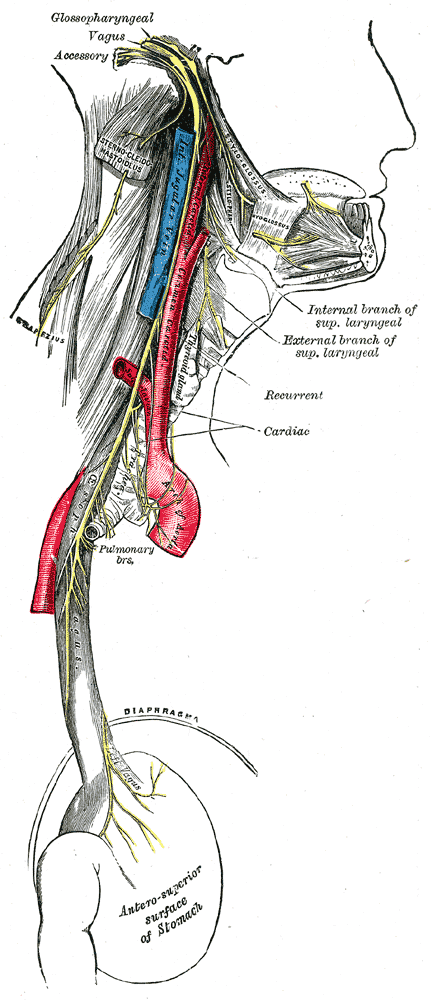
Glossopharyngeal Nerve
The nerves which originate directly from the brain are known as cranial nerves, as they originate inside the cranium. They are 24 in number (12 pairs), and their area of supply is restricted to the head and neck region except for the vagus nerve, which not only innervates the structures in the head and neck region but also supplies different structures in the chest as well as abdominal cavities. The glossopharyngeal nerve is the ninth cranial nerve. It transmits different types of sensory information from the posterior part of the oral cavity and tongue, as well as it provides motor stimulation to one of the pharyngeal muscles. It also contains parasympathetic fibers, which carry impulses from the superior salivatory nucleus. The glossopharyngeal nerve is an important nerve that takes part in regulating the pressure of the blood as well as its chemical composition.
Read more about the Brain Stem
Origin and Course of the Glossopharyngeal Nerve
The glossopharyngeal nerve originates from the upper part of the medulla oblongata at its anterior aspect behind the olives (bilateral bulged structures on the anterolateral side of the medulla that represent the inferior olivary nuclei). It runs in the posterior cranial fossa and reaches the jugular foramen. It leaves the cranial cavity via the jugular foramen along with the internal jugular vein, vagus nerve, and hypoglossal nerve. It descends down and reaches the border of the stylopharyngeus muscle, moves forward, and passes between the superior and middle constrictor muscles of the pharynx, it comes to the oral cavity to supply the mucosa of the oropharynx and posterior one-third of the tongue.
Classification and Functional Components
The glossopharyngeal nerve is a mixed nerve. It is one of two cranial nerves that contain the highest diversity of fibers, i.e., 5 different types of fibers. The other nerve is the vagus nerve. The glossopharyngeal nerve contains special visceral efferents that provide motor information to a muscle of the pharynx. It also contains general somatic afferents that carry pressure and chemical sensation from the carotid sinus and carotid body, special visceral afferents that carry taste sensations, general visceral afferents that carry general sensations, i.e., touch, pain, temperature, and pressure, etc. The other functional component of the glossopharyngeal nerve is the general visceral efferents that provide parasympathetic innervation to the parotid salivary gland.
Nuclei of the Glossopharyngeal Nerve
The nuclei of cranial nerves are the collection of cell bodies of neurons forming that cranial nerve. There are three types of nuclei of the glossopharyngeal nerve. These include the main motor nucleus, sensory nucleus, and parasympathetic nucleus (inferior salivatory nucleus). These nuclei are briefly discussed below:
Main Motor Nucleus
This nucleus is present deep in the reticular formation of the medulla oblongata. It receives corticonuclear fibers from both cerebral hemispheres. Efferent fibers from this nucleus take motor impulses to the stylopharyngeus muscle, one of the muscles of the pharynx.
Sensory Nucleus
The nucleus of tractus solitarius is an important nucleus in the brain. A part of it forms the sensory nucleus of the glossopharyngeal nerve. The nucleus of the tractus solitarius is also associated with other cranial nerves that carry taste sensations. This nucleus receives taste information from the posterior one-third of the tongue via the central processes of the neuronal cells of the ganglion of the glossopharyngeal nerve. The second-order neurons ascend upward to the thalamus, which is the main sensory collection center of the brain. Third-order neurons from the thalamus take the taste information through the internal capsule and corona radiata to the lower part of the post-central gyrus.
The nucleus of the tractus solitarius also receives information regarding blood pressure via the general somatic afferents from the carotid sinus. This information is passed on to the dorsal vagus nucleus. This forms the afferent pathway of the baroreceptor reflex.
The fibers that take general sensations from the oropharynx, posterior one-third of the tongue, and from the middle ear, do not relay at any of these three nuclei but ascend upward and terminate in the spinal nucleus of the trigeminal nerve.
Parasympathetic Nucleus, Along with the NeuronalPathway to the Gland
The inferior salivatory nucleus receives fibers from the hypothalamic nuclei as well as from the nucleus of the tractus solitarius. It also receives olfactory information via the reticular formation of the brain. Fibers from the nucleus of the tractus solitarius and the olfactory pathway participate in the secretion of saliva in response to the good taste of the food. The presynaptic parasympathetic fibers run through the tympanic branch of the glossopharyngeal nerve. tympanic branch ends in the tympanic plexus, from where these fibers join the lesser petrosal nerve, which takes these fibers to the otic ganglion. From the otic ganglion, postsynaptic parasympathetic fibers innervate the parotid salivary gland.
Functions
Functions of the glossopharyngeal nerve are listed below:
Motor Functions
The glossopharyngeal nerve provides motor innervation to the stylopharyngeus muscle. This muscle originates from the styloid process and is inserted at the thyroid cartilage. It involves swallowing and elevates the pharynx and larynx.
Sensory Functions
The glossopharyngeal nerve takes the taste sensations from the posterior one-third of the tongue. Mostly the receptors that detect bitter taste are present in the posterior part of the tongue. It also carries general sensations like pain, temperature, pressure, touch, etc., from the posterior one-third of the tongue, oropharynx, and middle ear.
The carotid branch of the glossopharyngeal nerve takes information from the carotid body and carotid sinus. This plays an important role in the regulation of blood pressure (via baroreceptor reflex) and in the regulation of the composition of the arterial blood (via chemoreceptor reflex).
Parasympathetic Functions
The facial nerve supplies parasympathetic or secretomotor innervation to the parotid salivary gland. As mentioned earlier, Salivary glands secrete saliva, which is important for the digestion of food. Saliva also keeps the oral cavity moist and protects it from many oral infections as it contains protective agents like lysozymes and antibodies etc.
Glossopharyngeal Nerve Reflexes
Reflex actions associated with the glossopharyngeal nerve are discussed below:
Carotid sinus Reflex (Baroreceptor Reflex)
The carotid sinus plays a role in the regulation of blood pressure. Neurons in the carotid sinus or baroreceptors are the stretch receptor variety of the mechanoreceptors. An increase in blood pressure stimulates the baroreceptors, and they fire neuronal impulses. These impulses are carried by the carotid branch of the glossopharyngeal nerve to the solitary nucleus. Efferents from the solitary nucleus go to the vagal nuclei and the vasomotor centers. Vasomotor centers are inhibited, resulting in dilatation of the blood vessels. Vagal stimulation slows down the heart rate. Both of these effects lead to a decrease in blood pressure. This reflex can be initiated by massaging skin over the carotid artery.
Chemoreceptor Reflex
The carotid body is a neuronal cluster located at the bifurcation of the common carotid artery. It contains specialized neurons called chemoreceptors that sense the chemical composition of the arterial blood and help to regulate it, i.e., pH regulation. These chemoreceptors are specifically to the partial pressure of oxygen in the arterial blood. If oxygen tension in the blood drops, this leads to a decrease in blood pH. Chemoreceptors are activated, and impulses are sent to the medulla oblongata through the carotid branch of the glossopharyngeal nerve. The reflex pathway will stimulate the medullary respiratory centers, which increases the respiratory rate to get rid of the excess carbon dioxide.
Gag Reflex
When the posterior wall of the pharynx is stimulated by the spatula or even with a figure, there is a contraction of the muscles, and the pharyngeal wall gets elevated, or in other words, the subject gags. This is known as the gag reflex. The afferent pathway of this reflex is formed by the glossopharyngeal nerve, which takes the touch information to the medulla. In the medulla, the impulse is passed on to the motor nucleus of the vagus nerve. The Vagus nerve forms the efferent pathway of the gag reflex.
A similar reflex is a palatal reflex. When the soft palate is stimulated, it reflexively contracts and gets elevated. The afferent and efferent pathways for this reflex are the same as for the gag reflex.
Diseases of the Glossopharyngeal Nerve
There are many diseases that affect the nervous system. Some of them are systemic, which affect the nervous system as a whole, and some are localized to particular nerves. We will limit our discussion to the diseases of the glossopharyngeal nerve and lesions of the brain stem that affect the nuclei of the glossopharyngeal nerve.
Isolated lesions of the glossopharyngeal nerve are very rare. Usually, this nerve gets damaged along with the vagus nerve and hypoglossal nerve. In the case of lesions involving the glossopharyngeal nerve, there is loss of taste and general sensations from the posterior one-third of the tongue, absent gag and palatal reflexes, and no or little secretions from the parotid gland.
Damage to the glossopharyngeal nerve can be caused by neuritis, ischemic injury, trauma to the skull resulting in crush injury of the nerve, exposure to toxins or drugs, multiple sclerosis and diabetic neuropathy, etc.
Glossopharyngeal Neuralgia
It is a brief, severe, episodic pain in the throat, behind the mandibular angle, and in the ear. This pain can be triggered by swallowing etc. No specific cause for glossopharyngeal neuralgia is evident to date, however, there are some factors that can trigger the pain i.e., cough, sneezing, cold liquids swallowing and talking, etc. Treatment of this pain involves the use of analgesic drugs (i.e., NSAIDs and others) and surgery in which there is resection of the branches of the glossopharyngeal nerve involved in pain transmission.
Testing the Glossopharyngeal Nerve
General sensory afferents of the glossopharyngeal nerve can be tested by performing the gag reflex or palatal reflex. If there is a lesion of the glossopharyngeal nerve, these reflexes will be absent. The taste pathway can be checked by applying different tastes (especially bitter taste) on the posterior one-third of the tongue of the subject and asking him to recognize the taste. In the case of glossopharyngeal nerve lesions, there will be a loss of taste sensation from the posterior one-third of the tongue. The motor component of the glossopharyngeal nerve (that innervates the stylopharyngeus muscle) cannot be tested individually.
Lesions of the Brain Stem Affecting the Glossopharyngeal Nerve Nuclei
As the nuclei of the glossopharyngeal nerve are present in the medulla oblongata, lesions of the lower part of the brain stem may involve these nuclei. This results in signs and symptoms the same as in the case of damage to the glossopharyngeal nerve along its course. Raised intracranial pressure (RIP), as in the case of tumors of the brain in the posterior cranial fossa, may cause herniation of the brain stem through the foramen magnum. Pressure over the nuclei and traction of the cranial nerves results in paralysis of these nerves. The last four cranial nerves, glossopharyngeal, vagus, accessory, and hypoglossal nerves, are involved. The same happens in the case of Arnold-Chiari malformation, in which there is herniation of the cerebellar tonsils along with the medulla oblongata through the foramen magnum.
Summary
The glossopharyngeal nerve (CN IX) is the ninth cranial nerve. It is a mixed nerve and contains general visceral and sensory afferents, special visceral efferents, special visceral afferents, and general visceral efferents as its functional components. The glossopharyngeal nerve originates from the upper part of the medulla oblangata). It runs in the posterior cranial fossa and reaches the jugular foramen, leaving the skull via the jugular foramen along with the internal jugular vein, vagus nerve, and hypoglossal nerve. It descends down and reaches the border of the stylopharyngeus muscle, moves forward, and passes between the superior and middle constrictor muscles of the pharynx, it comes to the oral cavity to supply the mucosa and posterior one-third of the tongue.
The glossopharyngeal nerve has three nuclei, the main motor nucleus, sensory nucleus (nucleus of tractus solitarius), and parasympathetic nucleus (inferior salivatory nucleus). General visceral afferent fibers of the glossopharyngeal nerve go to the spinal nucleus of the trigeminal nerve. The sensory function includes the transmission of taste sensation from the posterior one-third of the tongue and general sensations from the oropharynx, middle ear, and posterior one-third of the tongue. The sensory function also includes the regulation of blood pressure and the oxygen tension of the arterial blood via the baroreceptor and chemoreceptor reflexes. Secretomotor supply is provided to the parotid salivary gland via the otic ganglion. Motor functions include innervation of the stylopharyngeus muscle. The glossopharyngeal nerve also forms the afferent pathway of the baroreceptor reflex, chemoreceptor reflex, gag reflex, and palatal reflex.
Diseases related to the glossopharyngeal nerve include glossopharyngeal neuralgia and other lesions. The cause of glossopharyngeal neuralgia is unknown, but the risk is increased with certain factors like cold liquids, swallowing, talking, coughing, etc. The glossopharyngeal nerve can be tested by the gag/palatal reflexes and by applying different tastes on the posterior part of the tongue. Damage to the glossopharyngeal nerve can be caused by neuritis, ischemic or traumatic injury, exposure to toxins or drugs and diabetic neuropathy, etc. Lesions of the brain stem (tumors of the brain stem, Arnold-Chiari malformation, etc.) involving the glossopharyngeal nerve nuclei result in the same signs and symptoms. Treatment involves the solution of the underlying cause by using either medications or surgical interventions.
References
Illustrated Anatomy of the Head and Neck, Fehrenbach and Herring, Elsevier, 2012
Blumenfeld H. Neuroanatomy Through Clinical Cases. Sinauer Associates, 2002
Ropper, AH, Brown RH. Victor’s Principles of Neurology, 8th ed. McGraw-Hill, 2005
Standring S (ed.) Gray’s Anatomy, 39th edition. Elsevier Churchill Livingstone, 2005Image Source: Glossopharyngeal Nerve