


Table of Contents
The Golgi tendon organ is an encapsulated sensory receptor through which muscle tendon fibers pass before getting attached to the bone. It is a mechanoreceptor that plays an important role in regulating muscle tone and protects the generation of excessive force and limits the possibility of fiber injury. They are found near the point of insertion not the origin, a condition most suitable to detect the tension in muscles.
Introduction to Golgi tendon organ
About 10 to 15 muscle fibers are connected to each Golgi tendon organ, and the organ is stimulated when this small group of muscle fibers is tensed by contracting or stretching the muscle. The Golgi tendon organ is different from muscle spindles as the muscle spindles detect mainly the length of muscle and changes in length while Golgi tendon organs detect the tension in muscle fibers. The tendon organ has both a dynamic response and a static response. When muscles are activated intensely, the muscle tension suddenly increases and the dynamic response is activated. However, within a fraction of a second firing of tendon organs settles to a lower level of steady-state firing that is almost directly proportional to the muscle tension which is termed the static response. Thus, Golgi tendon organs transmit information to the nervous system about the degree of tension and rate of change of tension at any instant.
Structure of the Golgi tendon organ
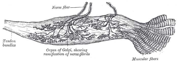
The Golgi tendon organ is a complex sensory receptor having a specialized bundle of muscle fibers linked to the muscle tendon, connective tissue, and neuronal endings. The muscle fibers are spindle-shaped and rich in contractile proteins while the surrounding connective tissue is continuous with the tendon. Connective tissue covering is responsible for protecting the Golgi tendon organ from damage and also provides structural support. It also helps to transfer the force of contraction to the Golgi tendon organs.
Neuronal connections of the Golgi tendon organ
Signals from the Golgi tendon organ are transmitted through large, rapidly conducting type Ib nerve fibers that average 16 micrometers in diameter, only slightly smaller than those from the primary endings of the muscle spindle. These fibers transmit signals into local integration areas of the spinal cord and, after making a synapse in the dorsal horn of the spinal cord, the signals are transmitted to the cerebellum through spinocerebellar tracts. Information also reaches to cerebral cortex through multiple neuronal connections. The local integration areas in the spinal cord excite a single inhibitory interneuron that inhibits the anterior motor neuron supplying the skeletal muscles. These local neuronal connections directly inhibit the individual muscle from having greater tension without affecting adjacent skeletal muscles.

Read more about Sensory Receptors
Location of the Golgi tendon organs
The Golgi tendon organs are found within tendons at the point where the muscle and tendon make the connection. This location allows the tendon organs to provide information about the tension generated by the contracting muscle, thus helping to regulate muscle tone and prevent injury. The Golgi tendon organs are most commonly found in tendons near joints and are distributed throughout the body, including the hands, feet, neck, and back.
Functions of the Golgi tendon organs
The Golgi tendon organs perform several crucial functions in the motor control of muscles. They prevent irregularities in muscle contractions and also modulate the contractile force.
Prevention of excessive tension in the muscle
If the muscle contraction is not regulated, it will go on unchecked and will tear away the fibers and tendons. It will also avulse the tendons from the bones. The Golgi tendons prevent the harm of these excessive muscle contractions. When it’s stimulated due to the excessive tension in tendons, signals are transferred to the spinal cord which produces inhibitory signals to stop the contraction. Due to these inhibitory signals, skeletal muscles relax and this phenomenon is called the lengthening reaction. As this reflex is inhibitory, it’s called the negative feedback control of muscle tension.
Inverse stretch reflex
The Golgi tendon organs execute the inverse stretch reflex. The harder a muscle is stretched, the stronger the reflex contraction of its fibers. However, upon contracting, when muscle tension becomes too much, contraction suddenly ceases to avoid injury. This relaxation in response to the strong stretch is termed autogenic inhibition or inverse stretch reflex. When a muscle is stretched, the receptor portion of the muscle spindles is stretched which excites its sensory nerve endings. These sensory nerve endings stimulate the integration centers of the spinal cord and in turn, stimulate the motor neurons to cause reflex contraction which is the stretch reflex. When muscle contracts tension is built up which stimulates the Golgi tendon organs. The Ib fibers from the Golgi tendons transmit this information to the local integration areas in the spinal cord. These areas trigger inhibitory postsynaptic membrane potentials and inhibit the motor neurons supplying the skeletal muscles. As a result, contraction ceases and tension in muscle tendons drops. This is called the inverse stretch reflex and is under the control of the Golgi tendon organs. The Golgi tendon fibers are in series with the skeletal muscle fibers and are subject to both active and passive stretch.
Equalization of the contractile force
The Golgi tendons equalize the contractile force of all the fibers present in muscle machinery and distribute the muscle load all over the fibers. They do so by inhibiting the muscle fibers generating excessive tension and stimulating the fibers with too little tension thus equalizing the contractile force. It protects the overloading of a few fibers or groups of fibers and maintains the uniform contractility of the muscles.
Regulation of muscle tone
The Golgi tendon organs are responsible for maintaining muscle tone. They adjust the contractility of individual fibers and prevent the individual fibers from either exceeding or falling down from the required tone. To adjust the tone, muscle spindles also play a role with the Golgi tendons to provide feedback information.
Postural stability
The Golgi tendon organs are responsible for maintaining posture. Specific postural control requires the programmed contraction and relaxation of the specific muscle groups. If any of the muscles involved in maintaining a specific posture is disturbed, postural control is lost. The Golgi tendon organs ensure that there is appropriate tension in the muscles making postural control possible.
Gait stability
Our walking pattern and gait are subjected to alteration by muscle tones and contractility. You can imagine the importance of proper muscular control for gait by observing an individual having cramps in a specific locomotor muscle. His gait and walking pattern will be greatly disturbed. The Golgi tendon organs ensure that all the muscles involved in locomotion and gait contract and generate enough tension to maintain normal mobility. If tendon receptors of the muscles at a joint are impaired, the control of proper mobility will be lost.
Medical conditions affecting the Golgi tendon organs
The Golgi tendon organs can be damaged in several medical anomalies. Here is a brief overview of the medical conditions affecting the Golgi tendon organs (GTOs) in muscles:
- Tendinitis is the inflammation of a tendon that causes pain and swelling. It may affect the functioning of GTOs.
- Tendinosis is the chronic degeneration of a tendon, typically caused by overuse or aging. It can impact the functioning of the GTOs. Tendinopathy is a general term used to describe any condition affecting tendons, including tendinitis and tendinosis.
- Muscle strain which is due to overstretched or torn muscle, can put pressure on the GTOs and cause pain or other symptoms.
- An injury to a ligament which is called a sprain can also affect the GTOs and cause pain or limited movement.
- Arthritis is the inflammation of the joints. It may be autoimmune, drug-induced, or other causes. It causes pain and limits the movement of joints, which can affect the GTOs.
- Overuse of muscles, tendons, and ligaments can lead to Golgi tendon damage and pain over time due to repetitive strain injury.
- Nerve entrapment or compression often caused by a herniated disk or other underlying condition, may occur. It can impact the neural tracts of Golgi tendon organs limiting their functioning.
- Bursitis is the inflammation of the fluid-filled sacs that cushion joints. It causes pain and limits the mobility of joints and can also affect the Golgi tendon organs in the vicinity.
It is important to consult a medical professional if you are experiencing symptoms or are concerned about your health. Diagnosing the abnormality in the beginning phase and sorting the necessary measures is the critical factor for a healthy and stress-free life.
How to assess the Golgi tendon organs clinically
The functioning of the Golgi tendon organs (GTOs) can be assessed in a clinical setting through various methods, including manual muscle testing, electrical stimulation, and imaging techniques.
Here is a brief overview of these methods:
Manual Muscle Testing (MMT)
MMT is a manual assessment of muscle strength, which involves applying force to a muscle while it is being contracted. It allows the physicians to assess the response of the Golgi tendon organs to the different levels of tension ranging from minimum to maximum. MMT provides valuable information about the functioning of the GTOs and the reflex pathways that control muscle contractility.
Electromyography (EMG)
EMG is a test that measures the electrical currents generated in a muscle. This test can be used to assess the functioning of the Golgi tendon organs by measuring the electrical activity in the muscle when it is contracted or stretched. The results of the EMG help the physicians to determine if the GTOs are functioning normally and providing appropriate feedback to the nervous system or not.
Nerve Conduction Studies (NCS)
NCS is a test that measures the speed and strength of nerve impulses. This test can be used to assess the function of the GTOs by measuring the electrical activity in the nerve fibers that are associated with the GTOs. NCS can provide important information about the function of the GTOs and the reflex pathways that control muscle tone.
Magnetic Resonance Imaging (MRI)
MRI is a non-invasive imaging technique that uses a strong magnetic field and radio waves to produce detailed images of the internal body structures. This test can be used to assess the function of the Golgi tendon organs by creating images of the tendons and ligaments that contain the GTOs. MRI provides information about the structure of the Golgi tendons and locates any structural abnormality compromising its function.
Ultrasonography
Ultrasonography is a non-invasive imaging technique that uses high-frequency sound waves to create images of the internal body organs. This test can be used to assess the function of the Golgi tendons by creating images of the tendons and ligaments containing the GTOs. It also helps to evaluate any structural abnormalities associated with muscles, tendons, ligaments, or GTOs.
These tests along with the symptoms and medical history of the patient help to diagnose the structural or functional abnormalities of the Golgi tendon organs and possible causes. Once the underlying cause is diagnosed, the physician devises a medical plan for treatment that may involve medication, physiotherapy, or surgery.
Summary
The Golgi tendons are the sensory receptors of muscles that are linked to the tendons. They comprise a few muscle fibers which are in series with the other fibers of skeletal muscles and are covered by the connective tissue.
They function to prevent damage to muscle fibers or tendons due to excessive tension in the muscles. Signals from the Golgi tendon organ are transmitted through large, rapidly conducting type Ib nerve fibers that average 16 micrometers in diameter. These fibers transmit signals into local integration areas of the spinal cord and, after making a synapse in the dorsal horn of the spinal cord, the signals are transmitted to the cerebellum through spinocerebellar tracts. The local integration centers of the spinal cord after processing the signals generate a response that is inhibitory in nature. These signals inhibit the motor neurons of the spinal cord and decrease the tension in muscles.
The Golgi tendon organs are located at the musculotendinous junction, mostly in the vicinity of the joints. They perform several functions such as limiting excessive tension, inducing inverse stretch response, maintaining muscle tone, equalizing the contractile strength of individual fibers, stabilizing the posture and gait, etc.
The inverse stretch reflex is a special phenomenon associated with the Golgi tendon organs. When a muscle is stretched, muscle spindles are activated which induces the reflex contraction of the muscle known as the stretch reflex. When this reflex contraction crosses a certain point, the Golgi tendons are activated which stimulates the integration centers of the spinal cord. The spinal cord, in turn, inhibits the muscles so relaxation occurs; this phenomenon is called the inverse stretch reflex.
Several medical conditions can affect the functioning of GTOs such as tendinitis, tendinosis, muscle strain, ligament sprain, arthritis, overuse of muscles, tendons, or ligaments, nerve entrapment or compression, bursitis, etc.
The function of GTOs can be assessed by manual muscle testing, electromyography, nerve conduction studies, magnetic resonance imaging, ultrasonography, etc. Medical tests and patients’ clinical history helps the physician to diagnose the underlying pathology affecting the GTOs and devise a treatment plan involving medication, physiotherapy, or surgery.