


Table of Contents
Cough is an important body response. We usually think of cough as a bad thing, but it is a protective measure adopted by the body. It is a warning sign indicating that there is something wrong with the airways. When there comes an irritant in the lower respiratory tract, a response takes place, and air in the lungs is expelled forcefully to get rid of that irritant. This happens again and again until the irritant gets removed or the cough is suppressed using medication. Cough is associated with many respiratory tract infections and may be productive (with sputum) or non-productive, depending on the disease. The vagus nerve forms the afferent part of the neuronal pathway involved in the cough reflex. In the following article, we will look at the vagus nerve’s origin, course, classification, and functional components. We shall discuss different nuclei related to it, its functions, its role in taste pathways, parasympathetic supply to different viscera, and different reflexes related to the vagus nerve. Last but not least, we shall have a look at the diseases caused by damage to the vagus nerve and the ways to check for its different lesions.
Vagus nerve
There are 12 pairs (24 total) of cranial nerves. These nerves provide innervation to the structures in the head and neck region of the body. The Vagus nerve is the 10th cranial nerve (CN X). It is a mixed nerve and supplies the structures in the above-mentioned region and different viscera in the chest and abdominal cavities. It is the longest cranial nerve. It provides motor innervation to the pharynx and larynx muscles (discussed later). Parasympathetic supply is provided via the vagus nerve to the heart, blood vessels, bronchi, part of the GI tract, and viscera like kidneys, liver, pancreas, etc. The Vagus nerve also transmits taste and general sensations to the CNS.
Read more about the Glossopharyngeal Nerve
Origin and course of the Vagus nerve
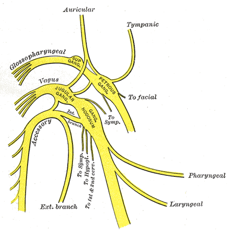
The Vagus nerve originates from the upper part of the medulla oblongata just below the origin of the glossopharyngeal nerve behind the olives (bi-lateral bulged structures on the anterolateral sides of the medulla that represent the inferior olivary nuclei). Like the other cranial nerves (except the trochlear nerve), it also emerges out at the anterior aspect of the medulla. It leaves the skull through the jugular foramen along with the internal jugular vein, glossopharyngeal nerve, and hypoglossal nerve and comes to the neck. It descends down in the neck within the carotid sheath. The Vagus nerve has two sensory ganglions; superior and inferior ganglions.
As many of the chest and abdominal cavity viscera are single (not in pairs like many other organs), the further route of each vagus nerve is different from the other. The right vagus nerve enters the mediastinum, passes behind the root of the right lung, descends down along the posterior aspect of the esophagus, and enters the abdomen via the esophageal opening of the diaphragm. Now it is called the posterior vagal trunk. Here, it divides into different branches, which take part in forming different plexuses.
The left vagus nerve enters the thoracic cavity, crosses the aortic arch, passes behind the root of the left lung, descends down along the anterior aspect of the esophagus, and enters the abdominal cavity via the esophageal opening. In the abdomen, it is called the anterior vagal trunk. It gives different branches which contribute to forming different plexus.
Classification and functional components
The Vagus nerve is a mixed nerve. It is one of two cranial nerves which contains the highest diversity of neuronal fibers, i.e., 5 different types of fibers. The other nerve is the glossopharyngeal nerve. The Vagus nerve contains special visceral efferents that provide motor information to the muscles of the pharynx and intrinsic muscles of the larynx. It also contains general somatic afferents that receive general sensations from the inferior pharynx and larynx, special visceral afferents that carry taste sensations from the root of the tongue and epiglottic taste buds, general visceral afferents that carry general sensations, i.e., touch, pain, temperature, and pressure, etc. from the thoracic and abdominal organs. Another functional component of the vagus nerve is the general visceral efferents that provide parasympathetic innervation to different viscera, i.e., the heart, etc.
Nuclei of Vagus nerve
The nuclei of cranial nerves are the collection of cell bodies of neurons forming that cranial nerve. There are three types of nuclei of the vagus nerve. These include the main motor nucleus, sensory nucleus, and parasympathetic nucleus (dorsal vagal nucleus). These nuclei are briefly discussed below:
Main motor Nucleus
This nucleus is present deep in the reticular formation of the medulla oblongata and also known as the nucleus ambiguus. It receives corticonuclear fibers from both cerebral hemispheres. Efferent fibers from this nucleus take motor impulses to the pharynx’s constrictors and the larynx’s intrinsic muscles.
Sensory Nucleus
A part of the nucleus of the tractus solitarius forms the sensory nucleus of the vagus nerve. The nucleus of the tractus solitarius is also associated with other cranial nerves that carry taste sensations. This nucleus receives taste information from the tongue’s root and the epiglottic taste buds via the central processes of the neuronal cells of the inferior ganglion of the vagus nerve. The second-order neurons ascend upward to the thalamus, the brain’s main sensory collection center. Third-order neurons from the thalamus take the taste information through the internal capsule and corona radiata to the lower part of the post-central gyrus.
The fibers that take general sensations from the root of the tongue, the lower part of the pharynx, and larynx and via the central processes of the neuronal cells of the superior ganglion of the vagus nerve, ascend upward and terminate in the spinal nucleus of the trigeminal nerve.
Parasympathetic Nucleus (Dorsal vagal nucleus)
The dorsal vagal nucleus is an important nucleus of the vagus nerve concerned with the parasympathetic supply to the smooth muscles of many thoracic and abdominal viscera. It is present in the medulla oblongata. Afferents to this nucleus come from the hypothalamus (autonomic control center) and also from the nucleus of tractus solitarius that afferent impulses of the carotid sinus reflex. Visceral afferents from the thoracic and abdominal viscera also form another afferent pathway for the dorsal vagal nucleus. Efferent from the dorsal vagal nucleus is distributed to the cardiac muscles and smooth muscles of the bronchi, esophagus, stomach, small intestine, and large intestine up to distal one-third of the transverse colon.
Functions
Functions of the vagus nerve are listed below:
Motor Functions
Motor innervation to the constrictor muscles of the pharynx. The superior and middle constrictors receive their nerve supply from the fibers of the cranial accessory nerve (CN XI) through the pharyngeal branch of the vagus nerve. Note that the accessory nerve’s cranial root is considered part of the vagus nerve. The inferior constrictor receives its nerve supply from the above-mentioned nerve and forms the external and recurrent laryngeal nerves, the branches of the vagus nerve. As the name shows, these muscles constrict the pharynx and are involved in the swallowing process.
The Vagus nerve supplies all the intrinsic muscles of the larynx via its recurrent laryngeal branch except the cricothyroid muscle, which is innervated by the external laryngeal branch of the vagus. These muscles are involved in phonation and voice production.
Sensory Functions
The Vagus nerve transmits taste sensations from the root of the tongue and epiglottic taste buds. It also carries somatic sensations (i.e., touch, pain pressure, etc.) from the tongue’s pharynx, larynx, and posteriormost part. Somatic afferents form the sensory pathway of the cough reflex. Visceral afferents carry sensations from the thoracic and abdominal organs like touch, pain, stretch, etc..
Parasympathetic Functions
One of the main functions of the vagus nerve is to carry the parasympathetic motor information to the different viscera like the heart, smooth muscles of the trachea, bronchi, stomach, small intestine, and large intestine up to distal one-third of the transverse colon. Vagal stimulation slows down the heart rate. In some instances of intense vagal stimulation, the heart may get stopped literally for a while. Stimulation to the smooth bronchial muscles causes bronchoconstriction. Vagal outflow to the gastrointestinal tract stimulates the GI smooth muscles and increases GI motility and secretions.
Vagus nerve Reflexes
Reflex actions associated with the vagus nerve are discussed below:
Carotid sinus Reflex (baroreceptor reflex)
An increase in blood pressure stimulates the baroreceptors in the carotid sinus, and they fire neuronal impulses. The glossopharyngeal nerve carries these impulses to the solitary nucleus. The vagus nerve forms the efferent pathway of the carotid sinus reflex. Fibers from the dorsal vagal nucleus take parasympathetic impulses to the cardiac muscles resulting in decreased heart rate. Efferents from the solitary nucleus also go to the vasomotor centers, and inhibiting them causes dilatation of the blood vessels. Both of these effects lead to a decrease in blood pressure.
Cough Reflex
An irritant in the lower respiratory tract can activate the receptors for the cough reflex. The vagus nerve forms the afferent part of the neuronal pathway involved in the cough reflex, through which impulses travel to the medulla oblongata. After processing, from the medulla oblongata, efferent impulses are transmitted via the motor nerves to the muscles, which contract, and air in the lungs is expelled forcefully to get rid of that irritant. This happens again and again until the irritant gets removed or the cough is suppressed using medication.
Palatal Reflex
When the soft palate is stimulated, it reflexively contracts and gets elevated. This is known as the gag reflex. The afferent pathway of this reflex is formed by the glossopharyngeal nerve, which takes the touch information to the medulla. In the medulla, the impulse is passed on to the motor nucleus of the vagus nerve. The Vagus nerve forms the efferent pathway of the palatal reflex.
A similar reflex is the gag reflex. When the posterior wall of the pharynx is stimulated by a spatula or even with a figure, the muscles contractions, and the pharyngeal wall gets elevated, or in other words, the subject gags. The afferent and efferent pathways for this reflex are the same as for the palatal reflex.
Diseases of the Vagus nerve
There are many diseases that affect the nervous system. Some of them are systemic, which affect the nervous system as a whole, and some are localized to particular nerves. We will limit our discussion to the diseases of the vagus nerve and lesions of the brain stem that affect the vagus nerve nuclei.
Damage to the vagus nerve can be caused by neuritis, ischemic injury, trauma to the skull resulting in crush injury of the nerve, exposure to toxins or drugs, multiple sclerosis and diabetic neuropathy, etc.
Hoarseness of voice
As discussed above, the vagus nerve supplies all the intrinsic muscles of the larynx. In case of lesions of the vagus nerve or damage to the recurrent laryngeal branch of the vagus nerve, any surgery in the neck area can lead to hoarseness of voice. In severe cases, there may be a completely absent voice.
Testing the Vagus nerve
Special visceral afferents of the vagus nerve can be tested by performing the gag reflex or palatal reflex. If there is a vagus nerve lesion, these reflexes will be absent. The motor component of the vagus nerve can be checked by asking the patient to speak something. If the subject cannot speak or there is hoarseness of the voice, vagal nerve palsy may be indicated.
Lesions of the brain stem affecting the Vagus nerve nuclei
As the nuclei of the vagus nerve are present in the medulla oblongata, lesions of the lower part of the brain stem may involve these nuclei. This results in signs and symptoms the same as in the case of damage to the vagus nerve along its course. Raised intracranial pressure (RIP), as in the case of tumors of the brain in the posterior cranial fossa, may cause herniation of the brain stem through the foramen magnum. Pressure over the nuclei and traction of the cranial nerves results in paralysis of these nerves. Mostly, the last four cranial nerves, glossopharyngeal, vagus, accessory, and hypoglossal nerves, are all involved in such lesions. Individual lesions are less common. The same happens in the case of Arnald-Chiari malformation, in which cerebellar tonsils are herniated along with medulla oblongata through the foramen magnum.
Summary
The Vagus nerve (CN IX) is the tenth cranial nerve. It is a mixed nerve and contains general visceral and sensory afferents, special visceral efferents, special visceral afferents, and general visceral efferents as its functional components. The Vagus nerve originates from the upper part of the medulla oblongata. It runs in the posterior cranial fossa and reaches the jugular foramen, leaving the skull via the jugular foramen along with the internal jugular vein, glossopharyngeal nerve, and hypoglossal nerve. The right vagus nerve enters the mediastinum, passes behind the root of the right lung, descends down along the esophagus posterior aspect, enters the abdomen via the esophageal opening of the diaphragm, and becomes the posterior vagal trunk. The left vagus nerve enters the thoracic cavity, crosses the aortic arch, passes behind the root of the left lung, passes along the anterior aspect of the esophagus, and reaches to abdominal cavity via the same opening.
The Vagus nerve has three nuclei, the main motor nucleus, sensory nucleus (nucleus of tractus solitarius), and parasympathetic nucleus (dorsal vagal nucleus). General visceral afferent fibers of the vagus nerve go to the spinal nucleus of the trigeminal nerve. The sensory function includes the transmission of taste sensation from the root of the tongue and general sensations from the lower part of the pharynx, larynx, and root of the tongue. Parasympathetic supply is provided to cardiac muscles and smooth muscles of the trachea, bronchi, GI tract, etc. Motor functions include innervation of all the intrinsic laryngeal muscles. The Vagus nerve also forms the efferent pathway of the carotid sinus reflex, cough reflex, gag reflex, and palatal reflex.
Diseases related to the vagus nerve include hoarseness of voice or complete voice absence. The Vagus nerve can be tested by the gag/palatal reflexes and by applying different tastes on the posterior part of the tongue. Damage to the vagus nerve can be caused by neuritis, ischemic or traumatic injury, exposure to toxins or drugs and diabetic neuropathy, etc. Lesions of the brain stem (tumors of the brain stem, Arnold-Chiari malformation, etc.) involving the vagus nerve nuclei result in the same signs and symptoms. Treatment involves the solution of the underlying cause by using either medications or surgical interventions.
References
Illustrated Anatomy of the Head and Neck, Fehrenbach and Herring, Elsevier, 2012
Blumenfeld H. Neuroanatomy Through Clinical Cases. Sinauer Associates, 2002
Ropper, AH, Brown RH. Victor’s Principles of Neurology, 8th ed. McGraw-Hill, 2005
Standring S (ed.) Gray’s Anatomy, 39th edition. Elsevier Churchill Livingstone, 2005Image source: Vagus Nerve