


Table of Contents
Mastication is a semi-automatic process. We can voluntarily control the movements of the lower jaw at the temporomandibular joint, but normally, during eating, the mastication process runs reflexively. When there is a food bolus between the upper and lower molar and pre-molar teeth, it stretches the masseter muscle. A stretch reflex is initiated, and the masseter contracts reflexively to elevate the lower jaw, grinding the food between the teeth. This continues until the food is completely ground and becomes ready for swallowing. A similar reflex can be initiated by stretching the masseter in other ways. This reflex is known as the mastication reflex or jaw jerk reflex. The neural pathway behind the jaw jerk reflex is the mandibular division of the trigeminal nerve. In the following article, we shall discuss the origin, course, classification, functional components, and branches of the mandibular nerve. We shall have a look at the nuclei related to the mandibular division in the brain stem, the mandibular sensory pathway and the trigeminal lemniscus, the function of the mandibular nerve, and the reflex(s) related to it. Lastly, we shall learn about some diseases related to the mandibular division of the trigeminal nerve.
Mandibular Nerve
In the head and neck region of the body, the peripheral nerve supply is made possible via cranial nerves, which directly originate from the brain, particularly from the brain stem in the case of most of the cranial nerves. These cranial nerves are 24 in number (in the form of twelve pairs). The trigeminal nerve is the 5th cranial nerve (CN V) and also the largest cranial nerve. It is the chief sensory nerve of the face area as well as mucous membranes of the mouth, nose and paranasal sinuses, etc. It also supplies motor innervation to the muscles of mastication. As its name suggests, it is made up of three big divisions named ophthalmic division (CN V1), maxillary division (CN V2), and mandibular division (CN V3). The ophthalmic and maxillary divisions are purely sensory, having no motor fibers, while the mandibular division is mixed in nature, containing motor fibers that supply the muscles of mastication as well as sensory fibers. Mandibular is the largest of the three divisions.
Classification and Functional Components
The trigeminal nerve is a mixed nerve and contains general somatic afferents and special visceral afferents. The first two divisions, i.e., ophthalmic and maxillary, contain only general somatic afferent fibers. The mandibular division contains both sensory afferents and visceral motor efferents.
Origin and Course of the Mandibular Nerve
The trigeminal nerve, the parent nerve of the mandibular nerve, arises from the brainstem at the anterior aspect of the pons in the posterior cranial fossa. It moves forwards in the middle cranial fossa lying over the petrous temporal bone and ends forming a crescent-shaped ganglion, called a trigeminal ganglion, present in the Meckel cave (a pouch formed by the dura mater). Arising from the trigeminal ganglion, the mandibular nerve (CN V3) passes forward in the middle cranial fossa and leaves the skull through the foramen ovale. Unlike the other two divisions, it does not enter into the lateral wall of the cavernous sinus (a big venous sinus that drains the brain). Outside the cranial cavity, it divides into several branches.
Branches of the Mandibular Nerve
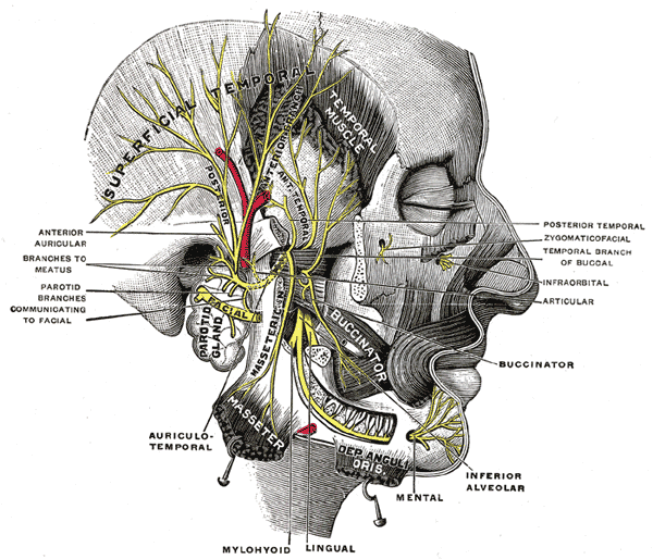
In the following words, there is a brief account of branches of the mandibular nerve. Two branches, named as meningeal branch and nerve to medial pterygoid muscle, originate directly from the mandibular nerve and innervate the associated structures. Then mandibular nerve divides into two trunks; anterior mandibular trunk and posterior mandibular trunk.
Anterior Mandibular Trunk
The anterior trunk is smaller and mainly contains motor fibers. This trunk gives rise to one sensory and three motor nerve branches:
Buccal Nerve
The buccal nerve is a small sensory branch of the anterior trunk of the mandibular nerve. it supplies the skin over the buccinator muscle and also the mucosa of the buccal region in the oral cavity. The name may confuse someone, but this nerve does not provide motor supply to the buccinator muscle. Motor supply to the buccinator comes via the facial nerve.
Nerve to Masseter:
This branch supplies the masseter muscle, an important muscle of mastication. This branch also supplies the temporomandibular joint. The nerve to the masseter is one that forms the efferent route for the jaw jerk reflex.
Nerve to Lateral Pterygoid
This branch supplies another muscle of mastication named lateral pterygoid muscle.
Deep Temporal Nerves
Deep temporal nerves supply the temporalis muscle, one of the muscles of mastication.
Posterior MandibularTrunk
This trunk is larger than the anterior trunk and mainly contains sensory fibers. It gives rise to:
Nerve to Mylohyoid
This nerve provides motor innervation to the mylohyoid muscle. This muscle forms the floor of the oral cavity.
Inferior Alveolar Nerve
The inferior alveolar nerve is sensory in nature. This nerve enters the mandibular canal via the mandibular foramen. It terminates by dividing into the incisive and mental branches. This nerve innervates the lower jaw and skin of the lower lip and gingiva. Distribution of different branches of this inferior alveolar nerve to different teeth is important during dental procedures such as tooth extraction.
Lingual Nerve
As the name shows, the Lingual nerve is the nerve of the tongue. This nerve receives the general sensations from the anterior two-thirds of the tongue. Remember that the facial nerve carries the taste sensations from the anterior two third of the tongue via its Corda tympani branch.
Auriculotemporal Nerve
Auriculotemporal is also sensory in nature, and it supplies the part of the auricle along with the tragus and temple. This nerve may get injured during parotid gland surgery.
Functions of the Mandibular Nerve
Sensory Functions
The mandibular nerve carries general sensations via general sensory afferents from the skin over the mandible, part of the cheek over the buccinator, skin of the side of the head, lower jaw along with part of the gingiva(gums), mucous membrane of the oral cavity and anterior two-thirds of the tongue. These sensations include pain, touch (light and crude), pressure, temperature (both warmth and cold), etc. It also transmits proprioceptive information from the muscles of mastication and the temporomandibular joint. The mandibular nerve does not carry any special sensation.
Motor Functions
The motor part of the mandibular nerve supplies the muscles of mastication. Muscles of mastication include masseter, temporalis medial pterygoid, and lateral pterygoid muscle. These muscles produce movements at the temporomandibular joint (the only moveable joint of the skull). These movements include protraction (forward motion of mandible), retraction (backward motion of mandible), elevation and depression of the mandible, and side-to-side rotatory movement of the mandible.
Nuclei of CN III Related to its Mandibular Division
The nuclei of cranial nerves are the collection of cell bodies of axons forming that cranial nerve. There are four nuclei of the trigeminal nerve. These include the main sensory nucleus, mesencephalic nucleus, spinal nucleus, and motor nucleus of the trigeminal nerve. All four nuclei collectively constitute the trigeminal system. These all are concerned with the mandibular division.
Main Sensory Nucleus
This nucleus is present in the pons. The sensations of touch and pressure are relayed here. The neuronal fibers coming here from the periphery have cell bodies in the trigeminal ganglion.
Mesencephalic Nucleus
The mesencephalic nucleus is present in the mid-brain and extends inferiorly to the main sensory nucleus. This nucleus receives proprioceptive information coming from the muscles of mastication via the mandibular division. These proprioceptive fibers have their cell bodies present in the mesencephalic nucleus and not in the trigeminal nucleus. Hence, they bypass the trigeminal ganglion.
Spinal Nucleus
It is in the form of a column extending from the main sensory nucleus, continuing through the medulla to the upper part of the spinal cord as far as the second cervical segment of the cord. The sensations of pain and temperature coming through the fibers of all three divisions of the trigeminal nerve are relayed here.
Motor Nucleus
This nucleus is exclusively related to the mandibular division of the trigeminal. Motor nuclei contain cell bodies of axons providing motor innervation to the muscles of mastication and some other. It receives impulses from the motor cortex via corticonuclear fibers, the tectum of the midbrain, the red nucleus, and the medial longitudinal fasciculus. It also receives fibers from the mesencephalic nucleus and forms a reflex pathway for muscles of mastication.
Mandibular Sensory Pathway and Trigeminal Lemniscus
General afferent fibers carry sensations from the area supplied by the mandibular nerve to the trigeminal nuclei. Carrier neurons at this step are first-order neurons. From the main sensory nucleus, spinal nucleus, and mesencephalic nucleus, the axons of the second-order neurons ascend upward in the form of a neuronal band or bundle, named trigeminal or spinal lemniscus, to the thalamus. Thalamus is the main sensory collection center of the brain. From the thalamus, sensory information is directed to the cerebral cortex. Axons of the third-order neurons reach the somatosensory area located in the post-central gyrus of the cerebral hemisphere, where all the general sensory information from the body is received, integrated, and used for making an appropriate and proper response by the motor cortex and also used for decision making and thinking process of the brain.
Jaw Jerk Reflex
The jaw jerk reflex is used to check the trigeminal nerve. When the lower jaw is stroked gently at the area just under the lower lip or at the chin when the mouth is slightly opened, the lower jaw reflexively moves upward to close the mouth. This is due to the stretch of the muscle spindles of the masseter muscle, which evokes the stretch reflex. The pathway for the jaw jerk is the mandibular division of the trigeminal nerve. The afferent information goes to the mesencephalic nucleus via the proprioceptive fibers of the mandibular nerve. Then it is sent to the motor nucleus of the trigeminal nerve. The impulse from the motor nucleus causes the masseter to contract to result in elevation of the mandible, i.e., closure of the mouth opening. A similar reflex occurs during the mastication process.
Diseases Related to the Mandibular Nerve and their Management
There are many diseases that affect the nervous system. Some of them are systemic, which affect the nervous system as a whole, and some are localized to particular nerves. We will limit our discussion to the mandibular nerve and its branches.
Trigeminal/Mandibular Nerve Palsy
Injury of the trigeminal nerve may result in paralysis of the muscles of mastication, resulting in problems with chewing and grinding of food when only the mandibular nerve is involved in the injury.
Causes of mandibular nerve palsy include diabetic neuropathy, aneurysm or tumor pressing the nerve, stenosis of the foramen from where the nerve leaves the skull, inflammation (neuritis), multiple sclerosis or vascular diseases resulting in ischemia, etc. Trauma to the head can result in crush injury of the nerve.
Trigeminal nerves can be clinically tested by a different method. The motor part of the trigeminal nerve is tested by asking the subject to clench his/her teeth. Failure to do so indicates mandibular nerve palsy. To check the sensory part, the cotton and pin method may be used. A piece of cotton is gently applied to the area supplied by the mandibular nerve. Lesions of the mandibular nerve result in loss of touch sensations. A blunt end pin is used to check pain sensations.
Trigeminal Neuralgia
Acute severe stabbing or burning pain on the face in the area supplied by the 2nd and 3rd divisions of the trigeminal nerve (maxillary nerve and mandibular nerve). This is also known as tic douloureux. The pain is episodic in nature and completely subsides after the acute attack is over. There is no facial muscle dysfunction or deformity. Usually, the pain is unilateral. The exact cause of trigeminal neuralgia is yet unknown. Pain can be triggered by even a slight stimulus like talking, chewing, or even a light touch on the face. Tic douloureux is one of the most painful conditions one can experience. Treatment involves both surgical and non-surgical interventions. In the surgical method, resection of one or more divisions of the trigeminal nerve is done. Pharmacological interventions include the use of NSAIDs and other analgesics.
Mandibular Nerve Block
Nerve block means anesthetizing a nerve providing analgesia as well as anesthesia. A trigeminal nerve block is a procedure used in routine clinical practice at dental clinics. Dental procedures such as tooth extraction from the lower jaw are done after the inferior alveolar nerve is anesthetized. Otherwise, the procedure is extremely painful. This block is also performed for the treatment of trigeminal neuralgia. This results in local anesthesia and analgesia in that area. In extremely painful conditions, a mandibular nerve block can be performed to relieve that pain, but this is a temporary remedy, and treatment of the underlying cause is required for the complete dissolution of the pain.
Summary
The mandibular nerve (CN V3) is the third division of the trigeminal nerve. It is a mixed nerve and contains general somatic afferents as well as general visceral afferents. The parent nerve, the Trigeminal nerve, emerges from the mid-brain at its anterior aspect at the level of the pons. It passes forward in the posterior cranial fossa, rests on the petrous temporal bone in the middle cranial fossa, and ends at a crescent-shaped ganglion, the trigeminal ganglion, located in Meckel cave. Here, it divides into three divisions; 1) Ophthalmic nerve (CN V1), 2) Maxillary nerve (CN V2), and 3) Mandibular division (CN V3). The mandibular nerve leaves the cranial cavity via the foramen ovale. It divides into several different branches; buccal nerve, nerve to lateral pterygoid, nerve to lateral pterygoid, a meningeal branch, nerve to the masseter, deep temporal nerves, nerve to the mylohyoid, lingual nerve, inferior alveolar nerve and auriculotemporal nerve. All four trigeminal nuclei are involved in the sensory and motor pathway of the mandibular nerve. The mandibular nerve provides sensory innervation to the skin of the cheek over the buccinator, the mucous membrane of the oral cavity and anterior two-thirds of the tongue, lower jaw (teeth and gingiva), lower lip, the skin of the side of the head, part of the auricle, tragus, and temple. Motor innervation is provided to the muscles of mastication and some other muscles, i.e., mylohyoid, anterior belly of digastric, etc. Sensations carried by the mandibular nerve include pain, touch, pressure, temperature, proprioception, etc. The mandibular nerve also forms the efferent pathway for the jaw jerk reflex. Diseases related to the mandibular nerve include trigeminal/mandibular nerve palsy and trigeminal neuralgia (most commonly, the maxillary and mandibular divisions are involved). Causes include inflammation (neuritis), pressure due to tumors, ischemic injury, traumatic injury, exposure to toxins or drugs, and demyelinating diseases like multiple sclerosis, diabetic neuropathy, etc. Lesions of the midbrain involving the spinal lemniscus may result in the same signs and symptoms. Treatment involves the solution of the underlying cause by using either medications or surgical interventions. A mandibular nerve block is commonly used in dental procedures (i.e., tooth extraction) and in the treatment of trigeminal neuralgia.
References
Illustrated Anatomy of the Head and Neck, Fehrenbach and Herring, Elsevier, 2012
Blumenfeld H. Neuroanatomy Through Clinical Cases. Sinauer Associates, 2002
Ropper, AH, Brown RH. Victor’s Principles of Neurology, 8th ed. McGraw-Hill, 2005
Standring S (ed.) Gray’s Anatomy, 39th edition. Elsevier Churchill Livingstone, 2005
Image source: Mandibular Nerve