


Table of Contents
Reflex actions are the rapid automatic and unconscious responses of the body to stimuli. They are a defense mechanism against injurious stimuli and play a vital role in maintaining homeostasis. Reflex actions are controlled primarily by the central nervous system, especially the spinal cord where they are integrated and executed without the involvement of a conscious segment of the brain. A classical example of reflex action is the withdrawal reflex. When your hand approaches the flame, it gets back without the conscious involvement of the brain to avoid injury. Later on, neuronal connections between the spinal cord and higher brain centers tell you about what has happened and make conscious apprehension of the situation. Reflex actions reflect your health and are used to diagnose various abnormalities by medical practitioners.
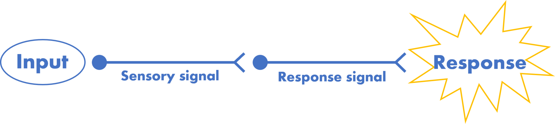
Reflex arc
The reflex arc is the pathway taken by a reflex action in the body. It involves the series of events that occur before a reflex action is produced. The basic component of reflex arc include:
- A receptor is mostly a sensory nerve ending that detects the stimulus and triggers afferent neurons.
- Afferent neurons transfer the sensory information from receptors to the integration centers of the central nervous system.
- The integration centers are mostly located in the spinal cord. They decide whether to produce a reflex action or not.
- Efferent neurons carry impulses from the spinal cord to the effectors to produce the response.
- Effectors are the organs that produce the response upon stimulation by the spinal cord. They are mostly smooth and skeletal muscles.

A reflex arc can be subjected to habituation. When an individual is continuously exposed to a stimulus for longer durations, the sensitivity of the reflex arc decreases so reflex action is diminished. The reflex arc can also be subjected to inhibition by conscious control. For example; an individual can hold his limb to a painful stimulus intentionally without eliciting a reflex response.
A reflex arc can be simple involving simple neuronal tracts or complex involving multiple neuronal tracts. Elbow jerk is an example of the simple reflex arc in which the tendon tap stimulates the stretch receptors (muscle spindles) and triggers the afferent neurons which carry information to integration centers that stimulate efferents to contract the biceps. In contrast, crossed extensor reflex which involves the extension of the contralateral limb when a painful stimulus is applied to one limb involves a complex reflex arc involving multiple neuronal tracts.
Classification of the reflex actions
Reflex actions are broadly classified into two types, somatic reflexes, and autonomic reflexes. Somatic reflexes involve skeletal muscles while autonomic reflexes involved smooth muscles and glands of the body.
Somatic reflexes
When you strike a tendon of a muscle with a hammer, it elicits its reflex action due to stretch activation of the muscle. It is a somatic reflex elicited by the somatic division of the nervous system. Somatic reflexes are numerous such as stretch reflex, withdrawal reflex, crossed extensor reflex, corneal reflex, etc.
Stretch reflex
The stretch reflex is the rapid contraction of skeletal muscle in response to stretch. It is one of the most basic forms of feedback control in the motor system. Stretch reflexes can also be called deep tendon reflexes and you can observe them easily by hitting the tendon of a muscle with a small reflex hammer.
When a skeletal muscle is stretched it depolarizes the muscle spindles as sodium channels open, causing an increase in the membrane potential. The stretch reflex maintains the normal muscle tone and prevents fiber damage as it limits the extent of stretch. It maintains normal muscular contractions, dampens oscillations, and stabilizes mobility. If there is no stretch reflex, muscle stretching will go unopposed tearing away the fibers. Stretch reflexes are commonly observed in clinical settings in various medical situations to know the normal working of the neuronal apparatus. In this procedure, the muscle tendon is gently tapped with a hammer, and a response is observed.
One of the most commonly observed reflexes for assessing motor function is the knee jerk reflex. When you tap the patellar tendon with a small reflex hammer, a leg jerk is produced due to the contraction of the quadriceps. It may be diminished or exaggerated or even absent under various medical conditions so it’s used as a diagnostic tool. Stretch reflex is exaggerated in several medical conditions such as:
- Hyperthyroidism
- Tumors or lesions of the spinal cord or nerve roots
- Vitamin deficiency, such as a deficiency of vitamin B1 (thiamine)
- Neurological disorders such as multiple sclerosis and peripheral neuropathy
- Degenerative diseases such as Parkinson’s disease
- Inflammatory conditions such as Guillain-Barre syndrome
- Increased intracranial pressure
- Upper motor neurons lesions, such as spinal cord injuries or tumors
- Hemispheric or brainstem lesions
- Chronic alcoholism
A diminished or absent stretch reflex is also an indication of neuronal or muscular abnormalities. Following are the conditions that decrease the responsiveness of muscles to stretch stimulus:
- Neuromuscular disorders such as muscular dystrophy and myasthenia gravis
- Peripheral nerve injuries or disorders
- Hypothyroidism
- Lower motor neurons lesions, such as poliomyelitis or peripheral nerve injury
- Vitamin deficiency, such as a deficiency of vitamin B12 (cobalamin)
- Chronic illnesses such as renal failure or liver disease
- Drug-induced conditions such as phenytoin (Dilantin) toxicity
- Inflammatory conditions such as polyradiculoneuropathy
- Degenerative conditions such as peripheral neuropathy in diabetes
- Chronic alcoholism
An exaggerated or diminished stretch reflex should be evaluated clinically to diagnose the underlying cause.
Withdrawal reflex
The withdrawal reflex is the rapid and automatic withdrawal of a limb or a side of the body from painful or injurious stimuli. The withdrawal reflex helps to protect the body from further injury and it’s totally an automatic response without involving any conscious part of the brain. However, neural connections between the reflex arc and the higher brain centers make you apprehend the situation. A common example of the withdrawal reflex is the withdrawal of your limb from flame or any such painful injury without your conscious perception.
Withdrawal reflex involves the common part of the reflex arc such as the receptor which detects the painful stimuli and sends impulses to the spinal cord which integrates the information and generates a motor response that triggers the effectors to produce the reflex. The withdrawal reflex is crucial to prevent further injury and the response produced depends upon the intensity of the stimulus, the location of the stimulus, and the sensitivity of the individual.
If the stimulus applied is a mild one, there will be a mild reflex such as a simple withdrawal of the limb however if an intense stimulus is applied, there will be an exaggerated response such as flinching or jumping. Abnormalities of the withdrawal reflex can indicate various medical abnormalities. For example, if withdrawal reflexes are absent it indicates peripheral nerve injuries such as peripheral neuropathy or nerve entrapment in various structures. An exaggerated withdrawal reflex indicates the increased level of sensitivity of the individual to the applied stimulus and it is a common symptom of hyperalgesia or allodynia. The withdrawal reflex is a common diagnostic tool in clinical examination. By assessing the withdrawal reflex healthcare practitioners can examine the integrity of your peripheral and central nervous system and can rule out any peripheral neuropathy. The withdrawal reflex is commonly tested by applying a painful stimulus such as a small little pin or a pinch to the skin and observing the response of the limb.
Crossed extensor reflex
Crossed extensor reflex is also elicited in response to painful or injurious stimuli. When an injurious or painful stimulus is applied to one limb, it flexes to move away from the stimulus, and the limb on the opposite side begins to extend. This phenomenon is called crossed extensor reflex. It functions to move the entire body away from damaging stimuli. Crossed extensor reflex is mostly observed after 0.2 to 0.5 seconds after the application of the painful stimulus. As it appears after a time-lapse, it involves multiple neuronal sets. Injurious stimulus is transferred from the afferents to the spinal cord where the information is interpreted and a flexor reflex of the limb is produced. Information is passed on in the spinal cord to the opposite side to control the opposite limb. Soon after, the opposite limb begins to extend to elicit a crossed extensor reflex. Crossed extensor reflex is also of great value in clinical settings. It is assessed by applying a painful stimulus to one limb and observing the response of the contralateral limb. Several medical conditions can either diminish or exaggerate the crossed extensor reflex indicating neuronal or muscular injury.
Corneal reflex
The corneal reflex also known as the blink reflex, is essential for protecting the eyes from harmful agents. It is a somatic reflex controlled by the 7th cranial nerve and provides valuable information about the working of neuronal apparatus. It involves the contraction of the orbicularis oculi muscle in response to the stimulation of the cornea by some external agent. It is a rapid and automatic response that prevents foreign objects, for example, dust or debris, from entering the eyes and damaging the cornea. The cornea is stimulated by the sensory fibers of the trigeminal nerve which send impulses to the integration center in the brainstem. Brainstem integration centers interpret all the information and stimulate the facial nerve to cause a blink reflex. The corneal reflex is tested during the clinical examination to assess the functioning of the facial nerve. To perform the test, a cotton swab is gently touched to the cornea which causes the eyes to blink. Corneal reflex proves a useful tool in conditions such as Bell’s palsy, brainstem strokes, or tumors.
Read more about Muscle Spindles
Autonomic reflexes
Autonomic reflexes are the unconscious automatic responses of the body to stimuli and the function to maintain the normal physiology of the body. These reflexes involve smooth muscles and glands of the body. They are elicited by the involvement of the autonomic nervous system which includes the sympathetic and parasympathetic nervous systems. Autonomic reflexes are of several types such as pupillary light reflex, cough reflex, baroreceptor reflex, and gastrocolic reflex. As these reflexes are elicited by the autonomic nervous system, they are subjected to alteration by autonomic drugs such as alpha-blockers or alpha agonists, beta-blockers or beta-agonists, etc.
Pupillary light reflex
The pupillary light reflex is an autonomic reflex that controls the size of the pupils in response to the light intensity. The reflex is under the control of the oculomotor nerve which stimulates the smooth muscle fiber controlling the pupillary size. When the eyes are exposed to bright light, the pupillary muscle constricts and reduces the amount of light entering the eyes. When pupils are exposed to dim light, pupillary muscles relax and the diameter of pupils increases to increase the amount of light entering the eyes. The pupillary light reflex plays a crucial role in maintaining visual function and protecting the eyes from damage by controlling the amount of light entering the eyes. The pupillary light reflex is useful to access the functioning of the oculomotor nerve and brain stem disorders. The conditions which affect the oculomotor nerve also affect the way pupils respond to the light. For example, anisocoria is a medical condition characterized by unequal pupils due to oculomotor nerve injury. The pupillary light reflex is also useful in assessing the activity of various drugs which either cause meiosis or mydriasis.
Baroreceptor reflex
Baroreceptor reflex functions mainly to regulate cardiac output and blood pressure. The baroreceptors are the specialized pressure sensors that are located in the walls of major blood vessels such as the aorta and carotid arteries. These receptors are capable of detecting the changes in blood pressure and transmitting this information to the central nervous system to take appropriate steps to maintain normal homeostasis. In response to the blood pressure changes, the baroreceptor reflex adjusts the activity of the sympathetic and parasympathetic nervous system so that cardiac output and vascular tone are regulated in a way to maintains normal blood pressure. When blood pressure is decreased, the baroreceptor reflex increases sympathetic tone to increase blood pressure. When blood pressure is decreased, the baroreceptor reflex increases parasympathetic activity to decrease blood pressure.
Cough reflex
The cough reflex is an important protective mechanism that keeps airways clear of mucous, dirt, or foreign particles. The cough reflex is initiated by the stimulation of specialized receptors in the airways which are known as cough receptors. Cough receptors when stimulated by various irritants such as mucus, or dust send impulses to medulla oblongata which integrates the information and stimulates the muscles of the abdominal wall, rib cage, and diaphragm to build up high pressure in the lungs which flows out and clears the airways.
Gastrocolic reflex
The gastrocolic reflex is a physiological response that occurs in response to the presence of food in the stomach. This reflex is responsible for stimulating the smooth muscle of the colon, leading to the evacuation of feces and the maintenance of normal bowel function. The gastrocolic reflex is initiated by the stretch receptors in the stomach which send signals to the integration centers in the CNS which stimulate the release of hormones and neurotransmitters increasing the motility of the colon. The gastrocolic reflex is an important component of the digestive system and helps to ensure the efficient elimination of waste products from the body.
Summary
- Reflexes are the automatic responses to a stimulus and are of great protective value. The neuronal pathways involved in eliciting a reflex action are called the reflex arc.
- They can be somatic or autonomic depending upon the division of the nervous system involved. If they occur in skeletal muscles controlled by the somatic nervous system, they are somatic reflexes; if they occur in smooth muscles and glands controlled by the autonomic nervous system, they are autonomic reflexes.
- Somatic reflexes include stretch reflexes, withdrawal reflexes, crossed extensor reflexes, corneal reflexes, etc.
- The stretch reflex is produced due to the stretching of muscle spindles. It prevents excessive muscle stretching and stabilizes contractility. The stretch reflex is elicited clinically by gently tapping the tendon with a small hammer which produces a jerk such as a knee jerk, elbow jerk, etc.
- Withdrawal reflexes involve withdrawing the limb or body part from a painful stimulus such as turning away the limb from flame unconsciously.
- Crossed extensor reflex is the phenomenon of extension of the contralateral limb on applying a painful stimulus to one limb.
- The corneal reflex is elicited in the form of blinking when a cotton piece s touched against the cornea. These reflexes are of great value to assess neural function.
- Autonomic reflexes include the pupillary light reflex which controls the amount of light entering the eye, the baroreceptors reflex which maintains the blood pressure, the cough reflex which clears the airways, and the gastrocolic reflex which evacuates the colon as a result of gastric stretching due to food.
References
Rodriguez-Beato FY, De Jesus O. Physiology, Deep Tendon Reflexes. [Updated 2022 Sep 11]. In: StatPearls [Internet]. Treasure Island (FL): StatPearls Publishing; 2022 Jan-. Available from: https://www.ncbi.nlm.nih.gov/books/NBK562238/